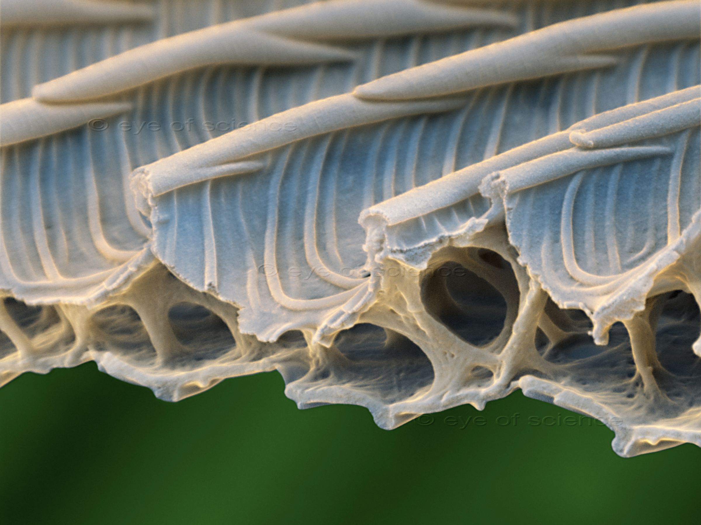
Butterfly Wing Scale
Section through a scale of a butterfly. These scales are hollow. Scanning Electron Microscope, 32000:1 (when 15cms wide)
Bruch durch die Schuppe eines Schmetterlings. Die Schuppen sind innen hohl. Raster-Elektronenmikroskop, 32000:1 (bei 15cm Bildbreite)

Jewel Wasp
The emerald cockroach wasp or jewel wasp (Ampulex compressa) is a solitary wasp laying its eggs on cockroaches. Scanning Electron Microscope, 25:1 (when 15cms wide)
Juwelwespe (Ampulex compressa), eine parasitär lebende Grabwespe, die ihre Eier auf Kakerlaken ablegt. Raster-Elektronenmikroskop, 25:1 (bei 15cm Bildbreite)
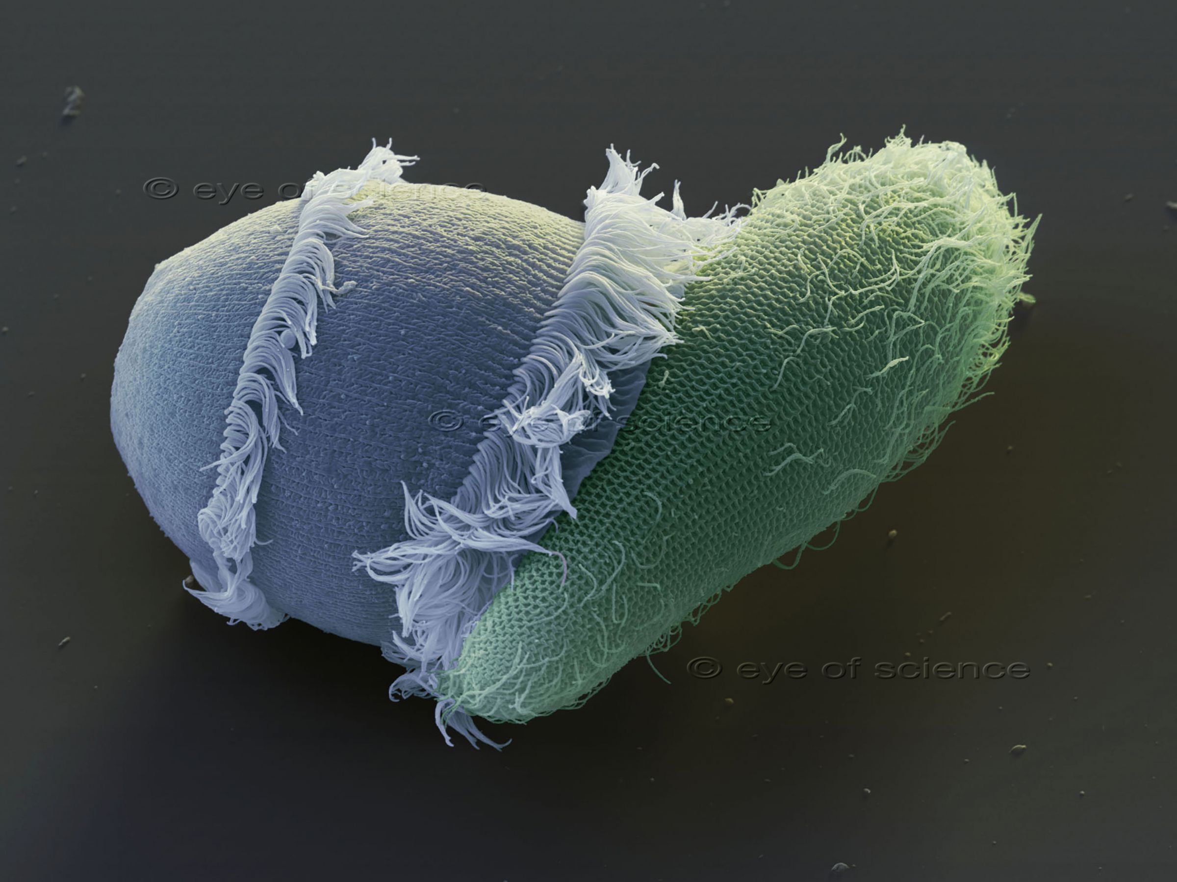
Predator- Ciliate
Didinium, a genus of ciliate mostly found in fresh and brackish water, attacking a paramecium ciliate. Scanning Electron Microscope, 730:1 (when 15cms wide)
Der Süsswasser-Einzeller Didinium beim Verzehr eines Paramecium (Pantoffeltierchen). Raster-Elektronenmikroskop, 730:1 (bei 15cm Bildbreite)
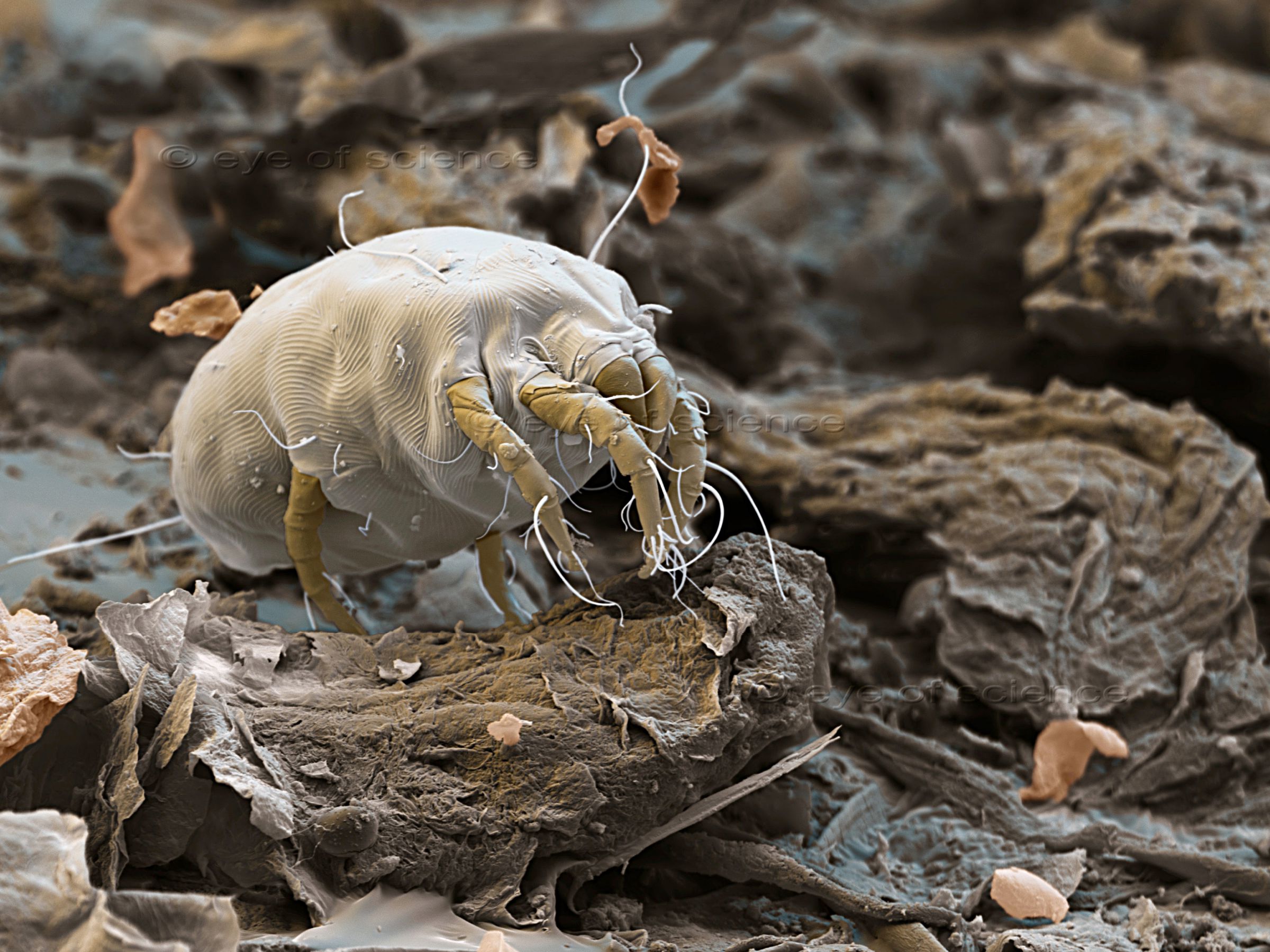
Dust Mite
Dermatophagoides house dust mite between skin scales. Scanning Electron Microscope, 400:1 (when 15cms wide)
Dermatophagoides Hausstaubmilbe zwischen Hautschuppen. Raster-Elektronenmikroskop, 400:1 (bei 15cm Bildbreite)

Skin of a Mite
Skin of the red mite Trombidium holosericeum. Scanning Electron Microscope, 1000:1 (when 15cms wide)
Haut der Roten Samtmilbe (Trombidium holosericeum). Raster-Elektronenmikroskop, 1000:1 (bei 15cm Bildbreite)

Wood Tick
Fully soaked Ixodes ricinus tick. Scanning Electron Microscope, 18:1 (when 15cms wide)
Vollgesogene Zecke Ixodes ricinus. Raster-Elektronenmikroskop, 18:1 (bei 15cm Bildbreite)
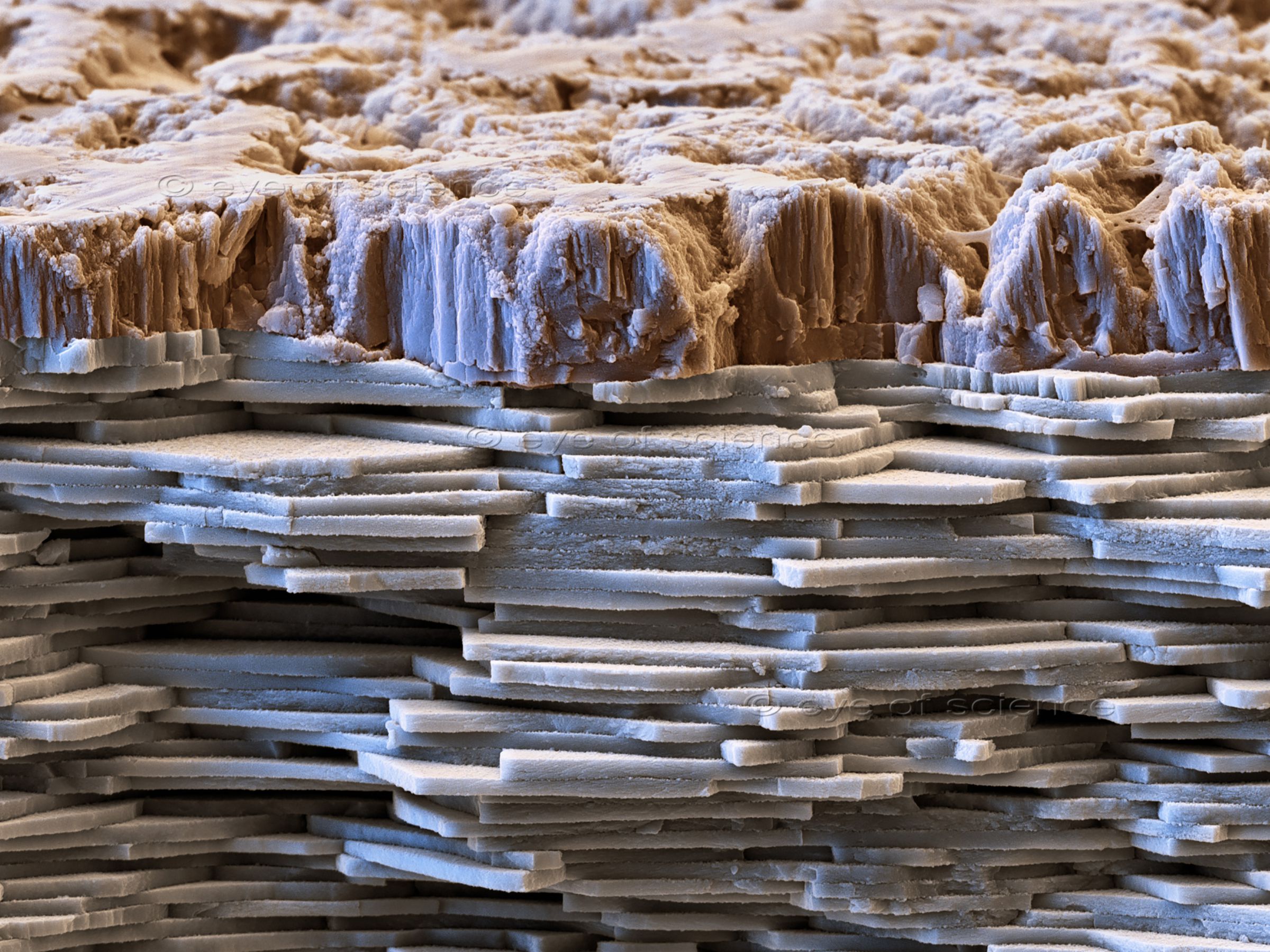
Nautilus Shell
Section through a nautilus shell, platelets of calciumcarbonate are visible. Scanning Electron Microscope, 5000:1 (when 15cms wide)
Bruch durch die Schale einer Nautilus, die Perlmutt-Plättchen sind gut zu erkennen. Raster-Elektronenmikroskop, 5000:1 (bei 15cm Bildbreite)
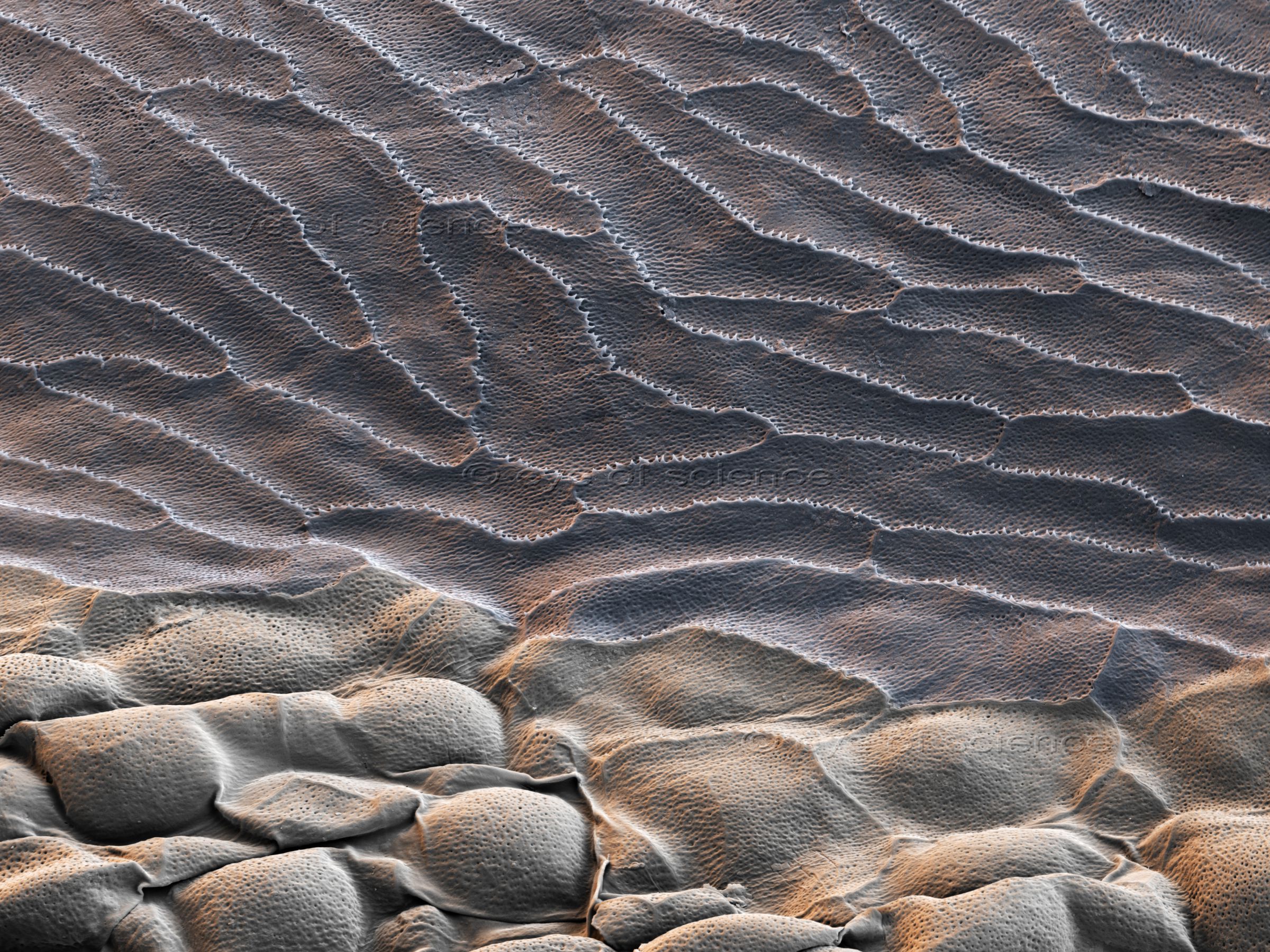
Snake Skin
Skin of a grass snake (Natrix natrix). Scanning Electron Microscope, 1200:1 (when 15cms wide)
Die Haut einer Ringelnatter (Natrix natrix). Raster-Elektronenmikroskop, 1200:1 (bei 15cm Bildbreite)
Wing of a butterfly with scales. Scanning Electron Microscope, 260:1 (when 15cms wide)
Der Flügel eines Schmetterlings mit Schuppen. Raster-Elektronenmikroskop, 260:1 (bei 15cm Bildbreite)
Butterfly
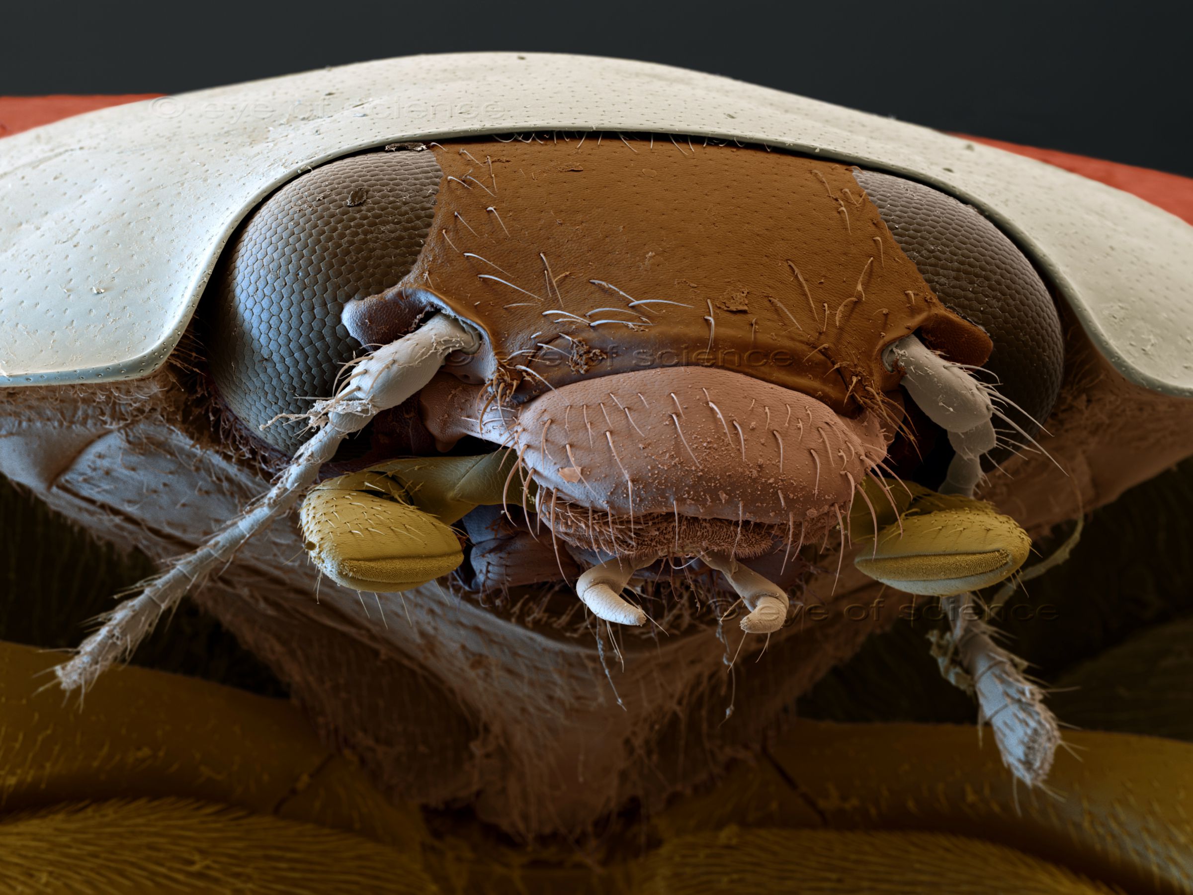
Ladybug
Portrait of an asian ladybug (Harmonia axyridis). Scanning Electron Microscope, 46:1 (when 15cms wide)
Portrait des asiatischen Marienkäfers (Harmonia axyridis). Raster-Elektronenmikroskop, 46:1 (bei 15cm Bildbreite)

Ant
Portrait of an ant with feeler, compound eyes and mandibles. Scanning Electron Microscope, 45:1 (when 15cms wide)
Portrait einer Ameise mit Fühlern, Facettaugen und Mandibeln. Raster-Elektronenmikroskop, 45:1 (bei 15cm Bildbreite)
Other topics
Andere Themenfelder
© Copyright for all images on this site by eye of science. All rights reserved.


